Video Otoscopy
Video Otoscopy is an essential tool in the treatment of many cases of otitis in dogs and cats and I have been using it daily for 15 years
 Video Otoscopy is an essential tool in the treatment of many cases of otitis in dogs and cats and I have been using it daily for 15 years.
The ability to clearly visualise the ear canal and, most importantly, the tympanic membrane and the facility to be able to accurately place flushing tubes, curettes and brushes means that a deep cleaning of the ear canal can be made in a safe manner.
One of the most common reasons for chronic otitis in dogs, other than the primary cause of atopy, in my experience, is the inability of home ear cleaning to thoroughly clear debris from the horizontal canal and many cases have firmly adherent material stuck to the tympanic membrane, which cannot be removed safely without the use of a video otoscope.
 Failure to remove this adherent debris can hinder the self-cleaning mechanism of the external ear called epithelial migration. I have seen many cases of recurrent otitis that have been cured purely by being able to finally do a proper deep clean of the ear canal.
If middle ear disease is present and myringotomy is necessary, this can only be done accurately and safely with the use of a video otoscope.
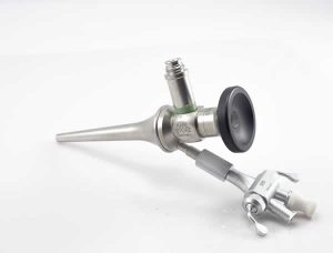
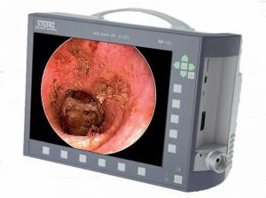
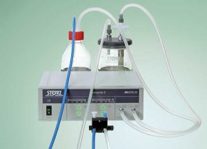 I have recently upgraded my Video Otoscopy set up with the new Storz Telepac Video Otoscope system with VetPump, which allows forceful but safe flushing and aspiration of debris from the ear canal. Previous to this, flushing was done with a syringe attached to the flushing tube, operated by a nurse.
I will shortly be also investing in a diode laser, where an optic fibre is passed through the channel of the video otoscope to allow me to ablate small tumours and areas of severe polypoid ceruminous gland hyperplasia or vaporise lesions of ceruminous gland adenomatosis in cats.
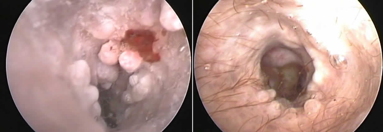
Polypid Ceruminous Gland Hyperplasia before and after treatment
Video Otoscopy also helps the pet owner to fully understand what is going on inside their pet’s ear and a record of the procedure is shown to them after every consultation and is provided for them on a memory stick if requested.
I will often also upload edited highlights of the procedure to my Vimeo account in selected cases and send links to the referring vet as a picture/video tells a thousand words!
Most veterinary ear referral cases will have at least one session of video otoscopy. The number of sessions required depends on the case, some cases of pseudomonas otitis requiring at least 3 or 4 sessions of video otoscopy and ear flushing.
If you wish to refer a case of otitis for Video Otoscopy, or any other veterinary dermatology case please ring 0116 3266759 or email referrals@dermvet.co.uk