LASER SURGERY
Two types of lasers are generally used in veterinary dermatology.
- CO2 laser, 10600nm wavelength– It has an absorption depth of less than 0.1mm and water is the main target molecule so it can be difficult to use via video otoscope where constant flushing with saline is necessary to keep a clear field of view. However, a recent paper has been published on the use of CO2 laser for treating proliferative otitis confined to the vertical canal only (Aslan et al, 2021). In October 2024 I purchased the Aesculight Vetscalpel CO2 laser and have used it successfully for ablating larger lesions in the ear canals such as tumours and larger Feline Cystamatosis lesions. The unit pushes air through the laser tip to remove any smoke/steam produced, to maintain a clear field of view.
- Diode laser, 810nm wavelength. This wavelength is heavily absorbed by melanin and less so by haemoglobin This is, in effect, thermocautery using concentrated infra-red laser light passed down an optical fibre, but is far more targeted, uses less power and less collateral damage than electrosurgery. It can be used within water and requires contact of the optical fibre tip with the tissue being ablated. The fibre can be passed down the working channel of a video otoscope. Despite now having a CO2 laser, I still find this useful for lasering smaller lesions with the horizontal canal.
CO2 lasers produce much less collateral thermal damage compared to a diode laser, but can be impractical for removing lesions very deep with the ear canal, where accuracy is essential to prevent inadvertent damage to the tympanum.
I am one of only two veterinary dermatologists in England and Wales with either a diode or CO2 laser which can be used via video otoscopy and I have saved many dogs and cats from needing TECA-LBO surgery using these wonderful machines
DIODE LASER SURGERY VIA VIDEO OTOSCOPY
There is little published in the veterinary literature specifically on diode laser use via video otoscopy. Gottelf (2019) has written a nice section in a recent textbook where he mentions both diode and C02 laser use (although he talks more about the CO2 laser) and in the same textbook Sobel (2019) has written on the use of diode lasers in minimally invasive and endoscopic small animal procedures including ears. There is also a recent case report by Hoshino et al (2022) of an aural polyp being removed by traction and then the base ablated with a diode laser.
There appears to be a lot more published on diode laser surgery in the human literature where it has been used in laryngeal surgery, turbinate surgery and in dentistry/orthodontics/oral surgery.
Whilst there are a number of different diode lasers on the market, one with a power of 15 watts is perfectly adequate for use within video otoscopy.
Equipment used
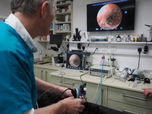
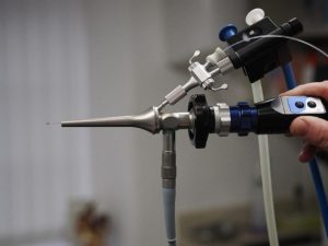
Karl Storz Telepack Vet X® with Video Otoscope and Vetpump II ® irrigation/suction system (Karl Storz Endoscopy (UK) Ltd)
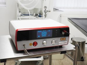
Angiomatics ® reconditioned Delta Laser 15W unit 810nm and 0.6mm Optical fibre.
The optical fibre is passed down the working channel of the Karl Storz otoscope. With the fantastic visualisation afforded by the Storz video otoscope it is possible to accurately place the tip of the optical fibre directly onto the lesion being treated. An in-contact method is used with the tip of the fibre in contact with the lesion being ablated. As this is done in a saline environment with constant flushing and suction, both the heat and smoke produced in the process is rapidly absorbed, minimising any thermal damage to surrounding tissue and obviating the need for expensive smoke evacuation equipment. I use a continuous setting at around 8-11 watts and use a footswitch to operate the laser, very often for split second at a time.
Since purchasing the laser in June 2020 I have used it to remove feline cystamosis lesions, polypoid ceruminous gland hyperplasia, tumours (ceruminous gland adenomas and adenocarcinomas) and inflammatory polyps. Several cases have had successful outcomes where previously TECA-LBO surgery would be indicated.
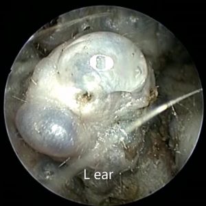
FELINE CYSTAMOSIS
Depending on the case and the extent of the lesions within the ear canal, between one and 3 sessions, spaced out at 2-4 weekly intervals to allow healing, may be necessary. Due to the risks thermal damage to adjacent, non-treated skin and the need to have adjacent skin re-epithelialize the wound, doing it this way minimised risk of non-healing or deeper, full thickness wounds in the ear canal. In between treatments, patients are sent home on steroid/antimicrobial ear drops. Power usually around 7 -10watts.
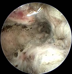
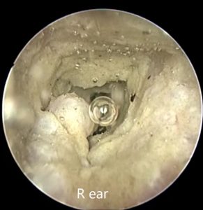
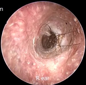
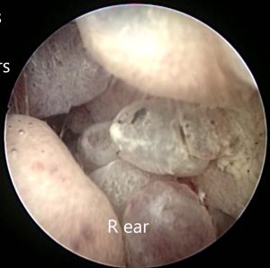
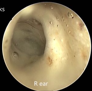
- Clyde, 7yo DSH Severe bilateral cystamosis
Clyde Severe Feline Cystamosis. Initial Presentation
Clyde. After 2 sessions with laser over 8 weeks
Clyde. After 3 sessions with laser, 11 weeks after initial presentation
To see video of Clyde, go to Video at https://vimeo.com/787283565
2. Lexie, 10 yo DSH Bilateral Cystamosis
Lexie. Feline Cystamosis (Cystadenomatosis) at initial presentation
Lexie. At 3rd and final session with laser, 7 weeks from initial presentation
To see Lexie’s video go to https://vimeo.com/787286255
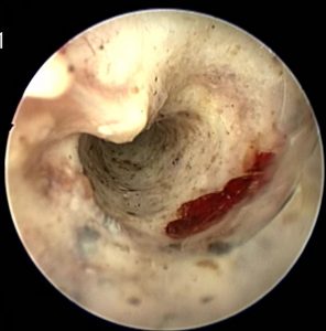
- Claude, 5yo DSH Mix of Cystamosis and inflammatory polyps
Clause. Mix of cystamosis and inflammatory polyps. At first presentation
Claude. After 10 weeks and 3 sessions with laser.
Video at https://vimeo.com/787328833
POLYPOID HYPERPLASIA
Similar management as with feline cystamosis. Between 1-3 sessions, spaced out at 3 weekly intervals may be necessary to remove all lesions safely. Patients sent home on steroid-antimicrobial ear drops after each session. Power 7-10 watts.
- Bo, 5 y.o. Welsh Springer Spaniel
Bo. Polypoid hyperplasia at presentation
Bo 3 weeks after laser surgery
Video at https://vimeo.com/787283415
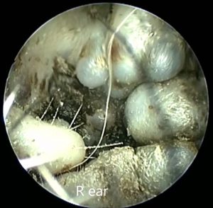
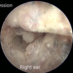
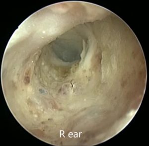
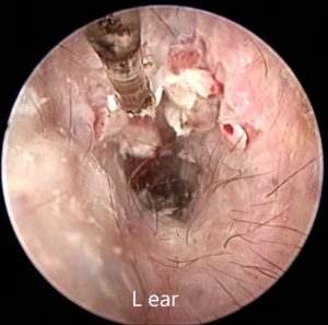
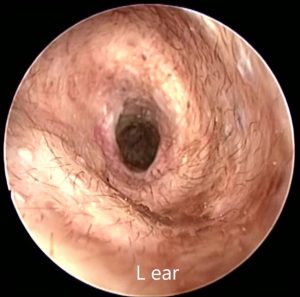
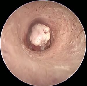
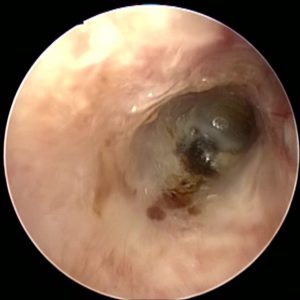
- Patch, 3 y.o. Shi Tzu Severe bilateral polypoid hyperplasia
Patch Right ear in case of bilateral severe polypoid hyperplasia.
Patch. Right ear after 16 weeks and 3 sessions with laser
Patch’s video at https://vimeo.com/787286693
- Lucy, 7y.o. Welsh Springer Spaniel
Lucy. Lasering polypoid hyperplasia
Lucy 6 weeks after lasering
Lucy’s video at https://vimeo.com/787286490
INFLAMATORY POLYPS AND TUMOURS
If these are large and accessible, they are removed by traction/avulsion using small, curved mosquito artery forceps and the remaining tumour/polyp tissue then ablated using the laser. If not accessible to traction the laser itself is used to break the mass down until the base is treated. Power between 7 and 11 watts depending on size and need to ablate large masses.
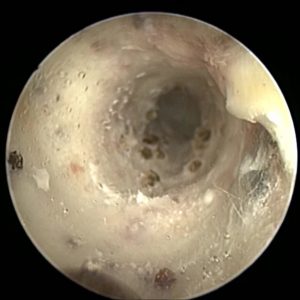
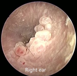
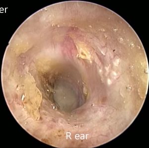
- Pepsi, 9 y.o. DSH Ceruminous gland tumours/cysts
Pepsi. Feline ceruminous cysts
Pepsi. 8 weeks after lasser
Pepsi’s video at https://vimeo.com/787348290
- Ruby, 10 y.o. DSH Ceruminous gland adenoma, with relapse and re-laser
Ruby. Ceruminous gland adenoma at presentation
Ruby. 5 weeks after laser
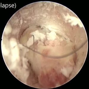
25 weeks after first laser.. regrowth of ceruminous gland adenoma
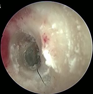
Ruby. 25 weeks after second laser. No regrowth of tumour.
Ruby’s video at https://vimeo.com/787287038
Myringotomy
A diode laser is a very fast effective way of performing a myringotomy. The site for a myringotomy is in the middle of the area between the manubrium of the malleus and the caudal edge of the pars tensa of the tympanic membrane. The power required is low and should not exceed 6 watts. Myringotomies performed by laser tend to heal a little more slowly than those done with sharp instrument.
Video at https://vimeo.com/787388263
COSTS
Every case is different and it depends on the extent and severity of the lesions and how many sessions are required to treat them all, but ranges between £800 – £4500 which may seem expensive, but is less so, very often, than doing TECA-LBO surgery and since 2020 I have saved many cats and dogs from having to have TECSA-LBO surgery using this laser.
References
Gotthelf LN (2019) Laser surgery procedures of the ear. Laser Surgery in Veterinary Medicine edited by Winkler C J Wiley Blackwell 102-115
Hoshino et al (2022) Diode laser‐assisted transcanal endoscopic removal of an aural polyp in the external auditory canal of a dog Vet Med Sci. 2022 8(5) 1862–1866.
Sobel DS (2019) Surgical lasers in minimally invasive and endoscopic small animal procedures. Laser Surgery in Veterinary Medicine edited by Winkler C J Wiley Blackwell 217- 237